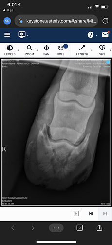
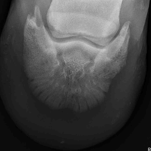
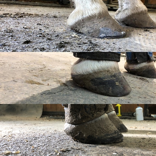
My life partner of a horse (the one in my avatar) was sound barefoot in boots and pads this spring until late June/ early July when she started to get touchy on the right front. I switched up her pads to something thicker which seemed to help for a while. Towards the end of July/ beginning of August she got worse so I took her in to a clinic for an emergency visit (a new to me clinic as the ones I normally use couldn’t fit her in). They did lateral views of all 4, used hoof testers and declared the only issue was thin soles and she needs shoes (along with several “it’s because she’s a thoroughbred” comments…).
Farrier comes out about a week later and we put flip flop pads on. Farrier notes an abscess on the medial side of the hoof is open and draining when applying the shoes. Two weeks go by without improvement, if anything she looks a bit more lame. Farrier comes out again to pull the shoe and see about floating the pad over the toe. When the shoe comes off, we see that the medial abscess is STILL draining (lots of nasty gunk) and there is now a second abscess on the lateral side. She also has some swelling in that same leg. We leave the shoe off, I wrap and pack the abscesses, do some PEMF on her RF and LH (as she is very sore bilaterally by now) and wrap all 4 with BOT. The next morning I rush her to our regular clinic for some more Xrays. Once we remove the hoof wrap at the clinic, we see 2 more abscesses have popped - one in each heel bulb (currently at 4 active spots in one hoof). The vet flushes those and digs a tract for the medial (which is going on 3-4 weeks now) and identifies two on the lateral side. Blood is drawn to test for inflammation markers to rule in/ out cellulitis which came back at 280 with 0-50 being normal. She had the radiographs looked over by the clinics surgeon who stated that they appeared normal. DX is to bute and give SMZs with stall rest and regular wrapping and packing of the hoof and 12 on 12 off of the leg with sweat wraps.
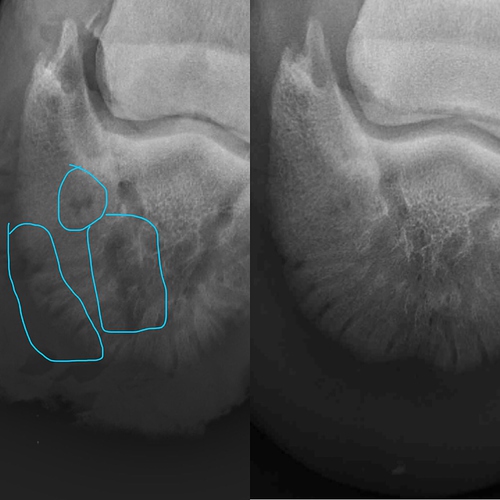
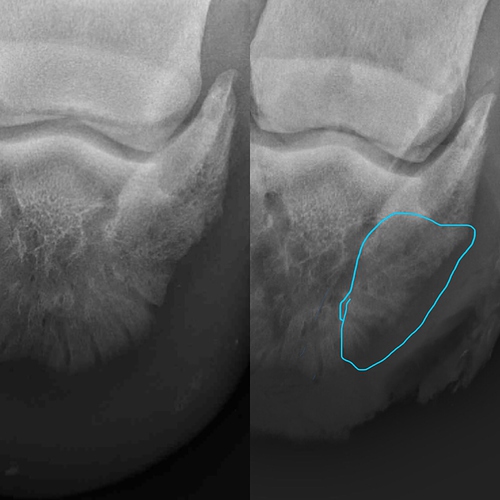
I was able to get a second opinion (roughly - they haven’t yet evaluated the horse, we go in for that Monday) who disagrees with the first surgeon and says there are significant changes and feathering of the bone between the radiographs from 2020 to the ones taken yesterday. They want me to bring her in for more imaging which we will do Monday.
To me, it appears we are headed down the same road we went down in 2016 when she suddenly became lame with an abscess that didn’t improve and was taken in to the second opinion vet above where they identified a bone infection in the coffin bone. She had surgery for debridement and a very long recovery time.
I am upset that vet 1 from early August brushed it off as thin soles and confused and worried that “history is repeating itself” with the same bizarre issue in the same foot. The surgeon at the second opinion clinic voiced a concern about that as well (will this be a continual thing? Why does it keep happening?). I appreciate any jingles, advice, commiseration. Radiographs below for those who are interested - should be current, 2020, side by side comparisons with the blue circles on the current xrays highlighting areas I am concerned about.

