Intermittent, mystery hoof lameness - is this soft tissue? - help

I would get him to your nearest referral center asap to make sure you aren’t dealing with an infection in that coffin joint. And if so, to start him on IV abx as well as get the joint flushed, etc.

This is a situation when a bone scan may also provide some help - if you have something going on further up the leg (eg, fractured scapula).

1 Like

You’ve gotten some excellent suggestions from other posters, but I just want to throw one other possibility out there. Intermittent lameness can be a symptom of lyme disease. It’s worth discussing with your vet, and it can be diagnosed via a relatively simple blood test.

Good luck.

6 Likes

Update:

Big guy has stayed lame for 24 hours finally. We started by having our farrier watch him go under saddle. He was significantly lame (3/5) tracking both directions, but felt worse when tracking to the right. Lameness was pretty much the same on straight lines and through the turns. Farrier said it looks RF to him, it felt LF to me under saddle.

We got off and pulled both shoes. After removing the impression material, we hoof tested him. He is reactive to hoof testing, on the inside heels of both front feet (and a little reactive on the RF outside heel area as well, but not LF outside heel).

Farrier says no abscess, We decide to pull the impression material, widen the wedge pad for more space and do magic cushion underneath.

We jog him on the lunge line after making the shoe adjustments and significantly better an hour later. Sound on soft ground tracking left on the circle. About a 1/5 on soft ground tracking right on soft ground circle (farrier says RF, I see LF). It’s very hot today, and farrier saw enough improvement in 60-90min to believe this is a pressure related issue with both heels, didn’t want to keep trotting him or try hard ground today - give him 2ish days and re-jog then.

Any other ideas on what this could be? I’m worried about soft tissue injuries at this point. Still waiting to get an appt with my vet to start with ultrasound…

Pics shows where he was hoof testing reactive ^^

There are so many things that this could be. Collateral ligament rutpure or sprain. Joint infection. Caudal heel pain (aka “navicular”). Your farrier isn’t doing him any favors … Get him to a qualified veterinary lameness specialist asap.

4 Likes

We have a call in for his regular performance vet (who manages all of his performance/lameness problems over the last 4 years - he is the vet who just did the coffin injections) … he is ISELP certified. We just haven’t gotten an appt since the lameness really started yesterday (Saturday) afternoon.

I am not even remotely close to an expert, in anything. But I also have a big horse, not stoic, and had some minor on off lameness in front that couldn’t really be pin pointed and would come and go with no discernable pattern. Eventually decided to pull shoes, and give her some Dr Green time and it’s been a massive positive change. She’s in full work, 8 months later and her hooves are so different in shape and function. I had shoes on all 4 for 4 years…again no expert! But for such a big boy, to have such owchy feet needing serious shoeing and sore heels (and again I am not trying to make an armchair diagnosis based on pics but he has such big hooves and what looks like a really contracted causal heel area) can he have some time barefoot? If there’s nothing else serious going on that gets diagnosed it might be worth a try. I used to scroll old forums every night trying to figure out how to help, and something as simple as pulling shoes ended up really helping. Took a few weeks for her to be comfortable but it’s been really really positive long term. Hope your guy finds a good solution and it works out!

6 Likes

If there is some kind of injury that can heal with turnout, that could be why horse heal, not necessarily going barefoot.
Cause and effect, if there is no clear diagnosis, makes it hard to say how things fare later and why.
Just turning out also can be lost time if something needed addressing, why pursuing a diagnosis is best.

1 Like

I had this sort of thing happen, and it took an MRI to diagnose. Even blocks were not that helpful. And actually we did both feet and the more sound one at the time looked worse on MRI.

1 Like

@IPEsq Yes, I have a feeling it might come down to that. As a 5 year old, he went from 17hh to 18hh he is now in like 4 months and he starting moving wonky and we sent him for an MRI then. MRI then showed a bunch of mild this and mild that in both front feet, so we chalked it up growing pains and and gave him 6 months off to grow into himself. Obviously, he’s fully grown now but I do remember how MRI’s can pinpoint every little thing and sometimes cloud a diagnosis. My vet is lameness specialist and a genius with an ultrasound, so I’m hoping he will get back to as soon as Monday rolls around and see the big guy this week.

What happened with your horse? Was it presenting similar to this?

One of the (oh so many) things that may be related to intermittent lameness is collateral ligament strain or other issues. Obvs there are a million things that can happen in the foot, so this is by no means diagnostic, but that “it looks to be one side and feels like the other” rings a bell when it’s medial collateral ligament.

But I’m inclined to think it’s more related to the heels, mostly because it’s almost always the heels and you should never look for a zebras when there’s a horse standing in front of you. But the collateral ligament is the more likely zebra in my experience

3 Likes

He never was abscess lame but he had intermittent lameness for a while. MRI showed that the DDFT was in bad shape near the navicular bones. Unfortunately, I lost him to colic the day the MRI results came back but the prognosis wasn’t great. My vet also dissected his feet post mortem and found adhesions and cartilage erosion as well.

Also adding this was a big horse too (though I don’t think he reached 18h…I quit measuring at 6yo)…I got him as a young looking 4 and he kept growing through his 8yo year. The lameness showed up at 9. And he never made it very far in sport despite having a lot of athleticism and some very successful close relatives because it was always something physical going on, mostly in the axial skeleton. His full, older brother is a hand shorter.

1 Like

Intermittent lameness can. E caused by suspensory issues.

1 Like

My intermittent front lameness horse with DDFT strain (and other supposedly minor things) based on MRI was ultimately fixed by turning him out sans shoes for six months and then a meticulous return to work in excellent footing while still living out 24/7 (and shod). Still lives out, still in excellent footing, now shod in Eponas. And I drive a lot to keep him in that situation. But he went from a horse we could first not keep sound while showing 3’ and under (roughly three years of being more out of work to in work) to one that we could not get sound (tried for 9 months) to one that is sound and doing the 1-1.05-m jumpers.

2 Likes

This sounds like it could be navicular caused by issues with the DDFT. It often presents with bone edema, and I know you said there was a “geyser” when he was injected. I’d also be concerned about how contracted those heels are. I would probably refer to MRI if the ultrasound doesn’t show anything, so the inside of the hoof capsule can be imaged.

With the availability of standing MRI for the foot these days, and the relatively reasonable cost, I’d likely go straight there at this point. In my experience, some of these weird on and off lameness issues are collateral ligament injuries in the hoof, or other things on the sides of the foot. The horse can travel pretty normally until the foot lands unevenly or turns and then there is a big shot of pain, which makes the horse fairly lame for a time.

3 Likes

Update -

On Sunday the farrier was out, and he was 3/5 Lame tracking both directions under saddle, lameness the same on straight lines/turns/circles. We removed the shoes, and hoof tested and he hoof tested reactive in both heels on both fronts. We removed the impression material, widen the wedge pad for more space and substituted it with magic cushion. Jogged him on the line and immediate improvement in both directions, sound tracking left and 1/5 tracking right.

Yesterday Afternoon - barn is closed on mondays, we come out yesterday for his vet appt and he jogs much much better again undersaddle. Took a few weird, “tranter” style steps right when we picked up the trot and then it was gone and he looked and felt like himself. Vet said he could see a few steps here and there, but it swaps from RF to LF and only shows itself randomly when the foot is on the outside. Vet is inclined to think its foot pain, related to the shoes since we saw such drastic improvement from making a shoe change 48 hours ago.

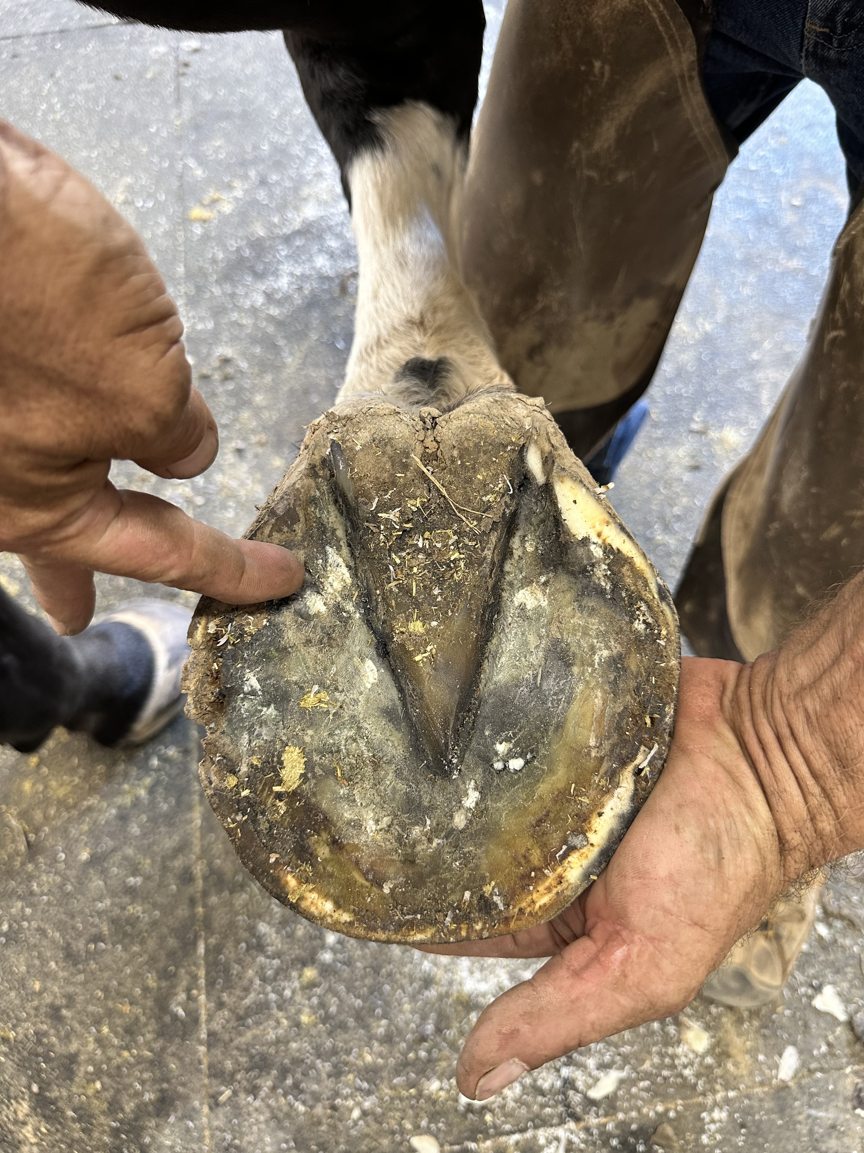
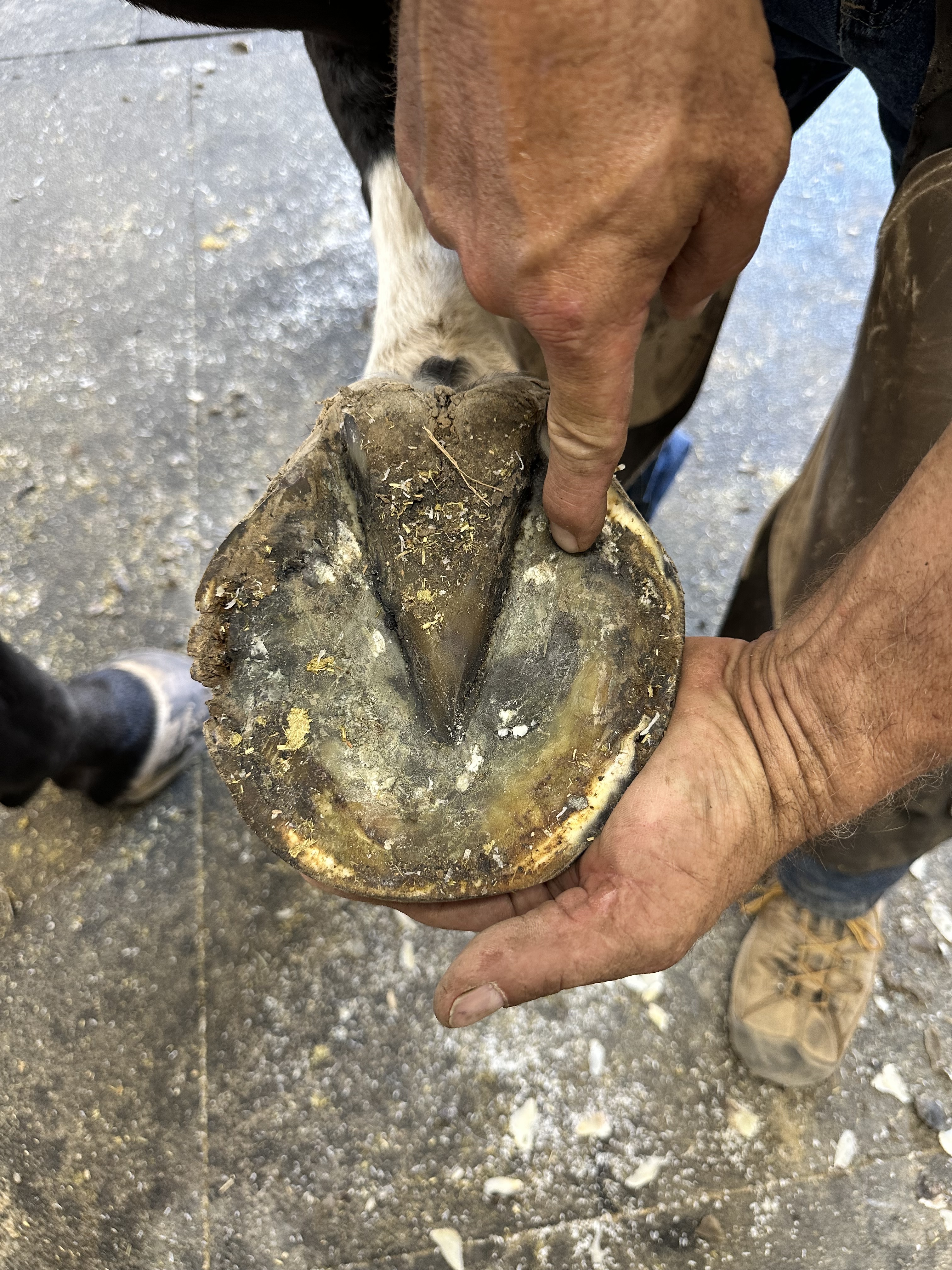
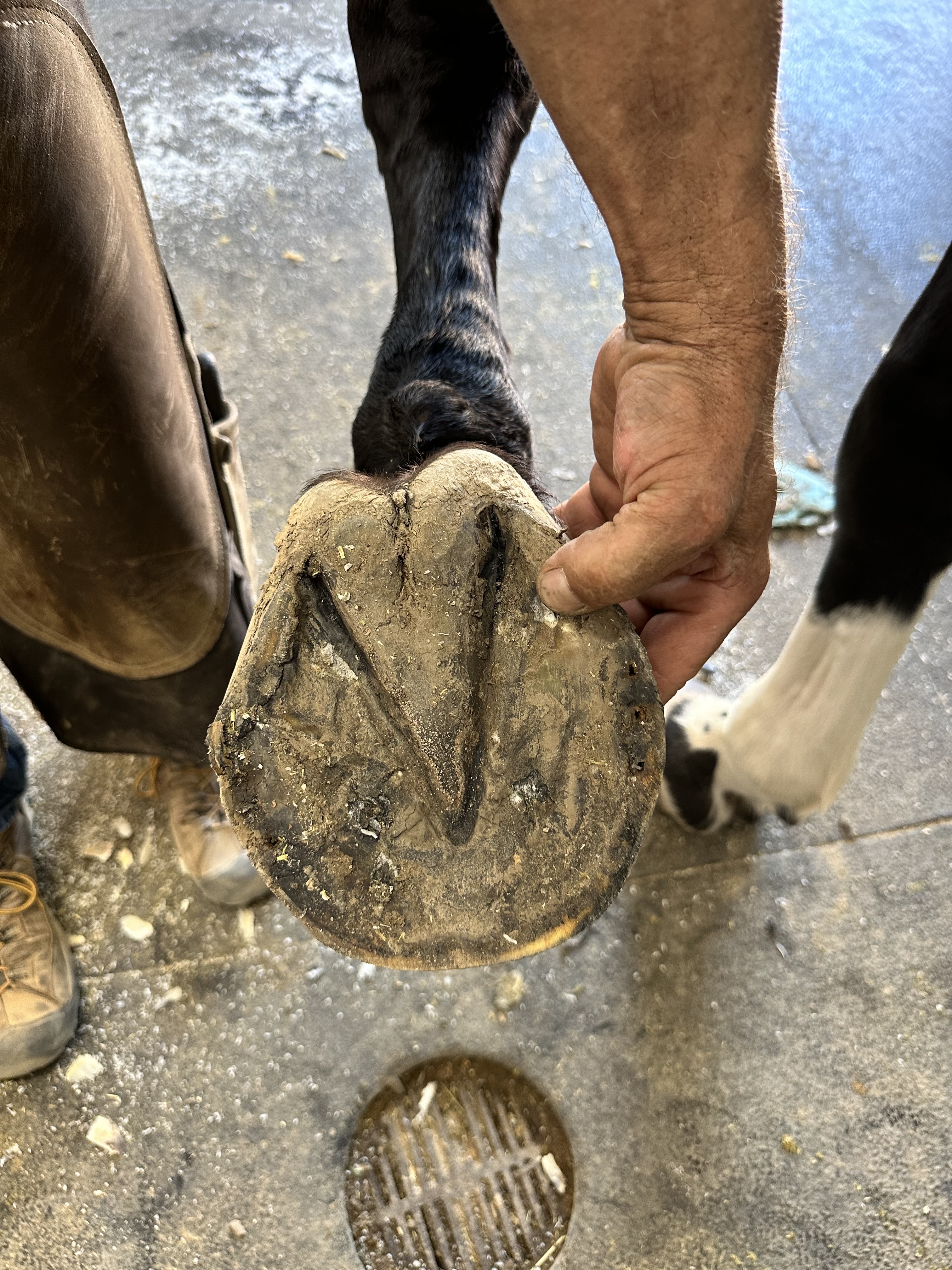
Next we go to X-rays, alot of x-rays. Vet is not pleased with current state of the feet. Apparently we have not balance x-rays since January, and things are different. His LF has a -1/-2 palmar angle, frog is wanting to prolapse (but hasnt yet), and heels are more contracted/crushed than before. His RF is a -1 palmar angle, frog is wanting to prolapse (but hasn’t yet), and heels are more contracted/crushed than before (this horse just had his feet redone 13 days ago). Next we pull the shoes, and snap more pics and coffin bone/navicular/etc all look fine.

Then we move onto Ultrasound. Using 4 different probes, we scan each front leg from the knee down. Suspensory, DDFT (we prepped the frog and were able to ultrasound through the frog to see insertion, coffin bone, navicular, etc. as well), proximal sesamoidan, lateral collaterals all look good. Medial Collaterals both look the same but don’t look the same as the lateral collaterals. We then pull-up his previous ultrasound scans of the Medial Collaterals from 2020, 2021, and 2022 and they look the same as the last scans. Fiber Patterns have remain unchanged, they just seem to be abnormally different but vet seems to think that with his history, this is who he is (he’s been sound and in work during all of those scans).

Vet believes based on his clinical presentation, shoeing and vet history…that the main problem is the shoes - he calls the farrier and they’re coming up with a new plan for his shoeing. They want a straight bar or a heart bar, possibly with a rocker toe or lateral roll and to go with a pour-in pad instead of impression material (but first we have to get the heel soreness out). Vet says don’t send for the MRI as he believes the medial collaterals are not clinical, he wants to fix the shoes first and go from there. I elect to shockwave the medial collaterals while we wait for the shoeing to be addressed. Farrier comes back out today to put shoes back on, and big guy is on hand walk/tack walk for 10-14 days while we figure out the shoes. Vet will come back and guide farrier with x-rays and emotional support through the next shoeing to get the angles and balance back under control.

5 Likes

No one else commented on the feet but I thought they are quite underun. His bony column support isn’t under his leg. I would definitely get good hoof balance X-rays and hopefully the farrier can get him back on the right track, foot wise.

Susan

4 Likes