Hip Surgery - Labral Tear/Impingement

If you look through my history I posted about my back a bit over a year ago. I had an MRI which showed a bulged disc, stenosis and facet joint arthritis. I had another back injection, targeted chiro and a lot of PT. To my surprise the PT really made a significant difference. I’ve been able to keep my pain really manageable with PT which is fantastic.

The downside is I started noticing a lot more pain in my hip. The PT also suspected a tear based on the back exam she did. Certain back PT exercises made my hip hurt so we had to modify my plan. I then got an MRI of my hip that showed a large labral tear on the right, a smaller one on the left and bone impingement on the right. To follow the order of insurance I had the hip injected. It provided some relief very short term, the Dr. was mainly using it to confirm the labral tear was causing the pain. Then I went through weeks of PT which just made the hip so much worse. We had to do the easiest exercises possible, decrease to once a week and finally got the all clear to stop thank god. My hip is now more painful and worse than before all of this but the diagnosis is clear and it’s more clear to me seeing how doing exercises with my left didn’t hurt at all how much that right hip is affecting me. I am fairly certain the original tear happened from a bad fall 20 years ago and it’s just progressing or re-tore recently.

So now it’s been recommended that I have the arthroscopic surgery to clean up the impingement and repair the tear. The after care and pain was more than I had expected and I’m a bit nervous. I can’t keep living like this riding is miserable, walking is miserable and believe it or not driving is the worst pain I have to deal with. I also now can’t sleep on that side at all. I’ve read some posts here where it seems like it’s a mixed bag if it helped or not. The posts I found were all from a few years ago. Anyone had this done recently? Any advice?

The Dr. I’ve seen is the hip specialist at a massive ortho practice near me that has great reviews and treats all of the professional sports teams around us. They focus on sports orthopedics which is why I chose them. The initial back Dr. wasn’t great with my hip so I switched to the hip specialist when all of this started. She’s done a lot of these surgeries and I really like her bedside manner which is rare in surgeons.

Labral repair is a hard recovery as the risk for reinjuring is high and healing takes a long time. I am currently scheduled for a total hip replacement (THR) in November and while scary, its a quicker recovery with a high success rate. Have you considered a THR?

My backstory: I had a major accident in 2001 and after years of horses and their antics, an MRI in January showed a torn labrum and a host of other things. 1st injection was miraculous and all was good until July. Injection in early August lasted until the anesthetic in the injection wore off, approximately 24 hours. Due to that second injection, I can not have surgery for 12 weeks and am struggling with sleeping, walking, sitting too long, you name it, it makes my hip mad.

Honestly, with that level of pain and the tear and the risk of re-injury as Spicy said, I would put for THR - total hip replacement

I had “significant arthritis” according to my surgeon. Bone on bone, several large spurs, with limited range of motion and increasing “stabbing” pain (like someone was literally jabbing a knife into my leg) with my leg giving out, by the time I had THR in March

I asked my surgeon if an MRI would be good to see if there were labral tears, etc, and he said don’t bother, even if there was, the bone issue was bad enough that if they had to go in there for labral repairs, they might as well go for THR

What do your hip joints look like? If there’s arthritis in there, rough edges, etc, they have a good chance of damaging the labrum even if surgery to repair the tears is successful, which was the biggest reason for me even checking to see the state of the labrum

@SpicyPRE you’ve got this! What approach will your surgeon do?

1 Like

I had a total hip replacement (left) coming up on 2 years ago. I was not a candidate for the labral tear repair as I had too much dysplasia.

I’m completely happy I did it. Pain instantly gone (and I mean instantly) and got my life back.

And just a little FYI - an MRI cannot “show” a labral tear (it doesn’t image cartilage). It can elude that a tear is there if the dye (hopefully they used that) gets into locations it is not supposed to be, but the only way to actually see the labral tear is when the surgeon is actually in the joint, looking at it. So MRI’s assume the tear is there … which it usually is.

1 Like

Choosing between arthroscopy and joint replacement is very rarely an option, because the arthritis that green lights joint replacement precludes arthroscopy. Surgeons are nearly always unwilling to replace a joint in a hip without severe arthritis.

It doesn’t sound like you have severe arthritis, so finding someone to replace the joint will be challenging to impossible. Nor should you go to that extreme procedure if the cartilage is intact. Amputation of the proximal end of the femur and replacing it and the acetabular cup with a prosthesis is still a Big Deal, and when things go wrong, the options aren’t awesome. A more conservative, preservative approach is far more appropriate when possible.

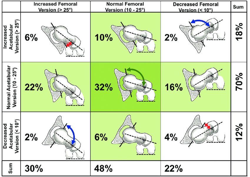
That said: labral repair is relatively “new.” We are still learning so much about who it helps and best approach, etc. So go get SEVERAL opinions, from the very best surgeons you can find. You may have to travel. Make sure you’re screened via pre op CT to evaluate version & acetabular coverage, because these things can impact your success. If there’s any indication that version or acetabular coverage are abnormal–and you should ask pointed questions, because many arthroscopists still consider these unimportant, despite recent research–you need to see a different surgeon who specializes in that type of anatomy, and that person is not an arthroscopist.

Labral repair has a high overall success rate. Make sure you’re screened for the things that we know can lower that. Make sure you see a surgeon that’s highly skilled, who does a shit ton of this surgery, who is staying current.

This isn’t correct. MRI at 3 Tesla is perfectly adequate to image labral tear, and most major centers are moving away from arthrograms.

If the exam is performed on a 1.5 Tesla machine, intra articular contrast is required, but it’s a resolution issue, not that the machine can’t “see” the labrum.

MRI is what DOES image cartilage.

2 Likes

Not sure about that. Being in a very active hip replacement group, nearly every situation has the surgeon saying “your quality of life takes precedence over how severe the arthritis is”, because even moderate arthritis can be in such a way that it’s causing major disruption. In general, they seem very focused on your level of pain, not simply what xrays do.

It’s the unfortunate few whose surgeons tell them they’re too young (despite severe arthritis) and/or that “your arthritis isn’t that bad, I won’t do the surgery” despite the person telling them their QOL is in the toilet.

Absolutely get a few different opinions on what’s going on, with a layout of the most to least conservative procedures and their pros and cons.

Anterior, muscle sparing approach. I am ready!

1 Like

This is really helpful thank you. On the MRI and x-rays no arthritis was mentioned. They did show me the images of the MRI in my consultation appointment after and it was pretty clear to see where the gap from a tear was in the right hip. I have not had a CT. Below are the findings from my xrays.

Joint spaces maintained.
Asphericity of the femoral head consistent with a CAM lesion
Right Alpha Angle: 60 degrees
Acetabular over coverage consistent with a PINCER lesion.

Right CEA: 37 degrees
Left CEA: 34 degrees

Right Tonnis: 0 degrees
Left Tonnis: 0 degrees

Medial joint space: 5 mm
Superior joint space: 5 mm
Lateral joint space: 5 mm

No acute bony fractures or aggressive lesions.
Soft tissues appear normal.

Result Narrative
AP Pelvis, Frog Lateral, Dunn

Does the Acetabular over coverage mean that I should see a different surgeon who doesn’t do arthroscopy? I have time to get additional opinions but I have a bit of a hard time knowing what questions to ask.

1 Like

Spare your leg from multiple surgeries and damage. Get the thr instead of the arthroscopic surgeries. The end result will be you tearing the fixed labrum and still needing to have thr.

Second opinion doesn’t hurt but you’ll probably hear the same info as the first surgeon provided.

1 Like

This is great that you’re at a center where the radiologist is evaluating center edge angle on labral tear imaging. These center edge angles are solidly in the normal range and not indicative of dysplasia. 0 degree tonnis supports that as well.

eta: there’s a great webpage here with a run down of this stuff, highly recommend: https://miles4hips.org/hip-dysplasia-whats-with-all-the-angles

I would still advise pursuing 3D CT to evaluate version. We think of these structures as two dimensional, but they exist in 3 dimensions and abnormalities in that, along with how they all relate to each other, can impact success. This is my fav image to illustrate version:

So we’re talking about the angle of the acetabulum & how that relates to the angle of the femoral neck & how that relates to the condyles in the knee. Variation here can absolutely cause instability, especially when you start removing some of the acetabular rim to address pincer FAI.

While Stryker hip mapping isn’t the only road to Rome (any good radiologist can do this) it’s a cool look at the data you get with CT:

https://microapp.stryker.com/hipmap/

With so much joint space noted and no signs of arthritis on the MR, joint replacement is absolutely not appropriate, and you would have a very difficult time finding a surgeon to agree to that procedure, if you did want to pursue it. It’s still a good question to ask (“do you see any signs here of arthritis, or are you worried about articular cartilage damage?”) as you work through your opinions, as surgeons and radiologists often disagree. But without arthritis, joint replacement is not (and should not be!) on the table. This is a good thing, you get to keep your parts!

Do go see a few more surgeons. Ask all your questions, including how many scopes they do in a year (you want someone doing 200+) and what their revision rate is and what’s driving their revisions. Ask how they’re opening and closing the capsule, because wow that varies a lot. And absolutely go with a surgeon who can communicate well and isn’t a dick–no one needs to settle for a surgeon who’s a dick.

Best of luck! It sounds like you’ve got things well in hand so far, and you’re definitely asking all the right questions!

3 Likes

Gotta tell you that I am jealous of your lack of arthritis.

1 Like

Thank you so much I really appreciate all of the info!! It was really funny getting the X-rays because the tech said ohhhh your Dr wants all of the weird angles let me go find her list lol

I’ll definitely meet a few other surgeons and I have a great list of questions now.

3 Likes

My surgeon told me that an MRI won’t directly image cartilage, and cannot actually see the tear, so that is why I repeated it here.

Perhaps based on the MRI available in my area at his disposal?

I’m not sure why he would say that, because it’s not true. Sounds like he was talking about a CT arthrogram, tbh.

I did mine in 2020 multiple falls from horses later I will say without hesitation that this was THE BEST choice I ever made. Do I have physical limitations now. Yes. But am I pain free and riding successfully. Yes! Feel free to pm me.

1 Like

Not sure, but that’s what he told me.
I had an MRI , not a CT. But I didn’t have any contrast dye used and they should have but I had enough changes on my xrays he didn’t need me to redo the MRI to “prove” what was going on because my xrays showed enough degradation of the joint space.

Even a 1.5 T MRI shows cartilage. It doesn’t have the resolution to show an acetabular labral tear without contrast but the cartilage appears. You can “see” it.

Here’s a paper that discusses how MRI visualizes cartilage. With pictures! So you can see cartilage, too.

The x ray based techs–radiographs, CT, fluoroscopy, etc–do not directly image cartilage. You make assumptions about the cartilage based on the space shown between the bones, or you use contrast.

I hope that’s … illuminating :grin::grin::grin:

1 Like