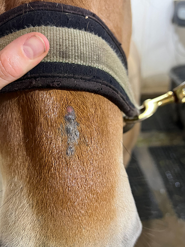
My Paint gelding had a cutaneous hemangiosarcoma removed from his belly when he was 26. It started out resembling an engorged tick but couldn’t be removed. I let it go, and it slowly developed into a dark pea-sized growth. Neither of our vets had ever seen anything like it, and one had retired a few months earlier after 50 years in practice.
When I brought him in one day it looked like he had scuffed it on something. As soon as I started to clean it up it started bleeding profusely, dripping all over the floor. I ran out of alumaspray. The grain store was out so I got a powered coagulant for goat horns that worked fine. The vet removed it with very large margins and it had not spread to muscle tissue. She sent it for biopsy. Apparently hemangiosacomas are seen in dogs and the dx was slanted a bit towards dogs. The lab wasn’t associated with a vet school so we sent it to Michinan State which confirmed the diagnosis as cutaneous hemangiosarcoma…
I also found an equine oncology specialist at Colorado State vet school who talked with my vet. Turns out her DVM is from there. The specialist said there was little to be concerned about. He said it was unlikely to reoccur and hadn’t spread because it was in the skin. I recall taking a day off from riding. The vet did a terrific job with the sutures. You could find the scar when he had his summer coat if you knew where to look.
This is the lab desciprtion. I’m including it because this literally was the size of a pea and had every known type of tissue and blood vessels known to horses. JB is probably the only one who will understand it all. 
Microscopic Description
Three sections representing the received haired skin specimen are examined. The dermis is expanded and effaced by a fairly well demarcated, unencapsulated, highly cellular, multinodular proliferation of neoplastic cells arranged in dense interlacing streams supported by moderate amounts of collagenous fibrovascular stroma. Neoplastic cells are spindloid with indistinct cell borders and contain small amounts of eosinophilic cytoplasm. Nuclei are oval, vesiculate and contain 1-2 variably prominent nucleoli. There is moderate anisokaryosis and 16 mitoses in 10 high-power fields (400x). There are numerous small, blood-filled clefts scattered throughout the mass. Small numbers of neutrophils and eosinophils are scattered throughout the mass and adjacent stroma. There are also large amounts of granulation tissue throughout the mass, particularly along the periphery and the overlying ulcerated surface. Such granulation tissue is composed of reactive fibroblasts, collagen bundles and perpendicularly oriented blood vessels. The overlying epidermis is extensively ulcerated and replaced by a dense layer of necrotic debris mixed with hemorrhage and large numbers of degenerate neutrophils. Regionally within the superficial dermis, there are moderate amounts of homogenous amorphous eosinophilic material, interpreted as sclerotic collagen. In one section, there is a tract-like lesion that extends from the surface into the mass that contains mild hemorrhage and is lined by granulation tissue and necrotic debris. Such granulation tissue raises the overlying skin surface, consistent with exuberant granulation tissue.
The findings are most consistent with a poorly differentiated sarcoma. Given the described blood-filled clefts, the primary differential is hemangiosarcoma.


 The one drug (Tagamet/cimetidine) we used to be able to get is off the market
  and if you do surgery or cryo on then on cats you just make the sarcoid angry and it redoubles efforts to take over as much of the affected area (usually nose