Unlimited access >

Recurring skin abscess

Already posted on horse vet corner and horse health discussion.

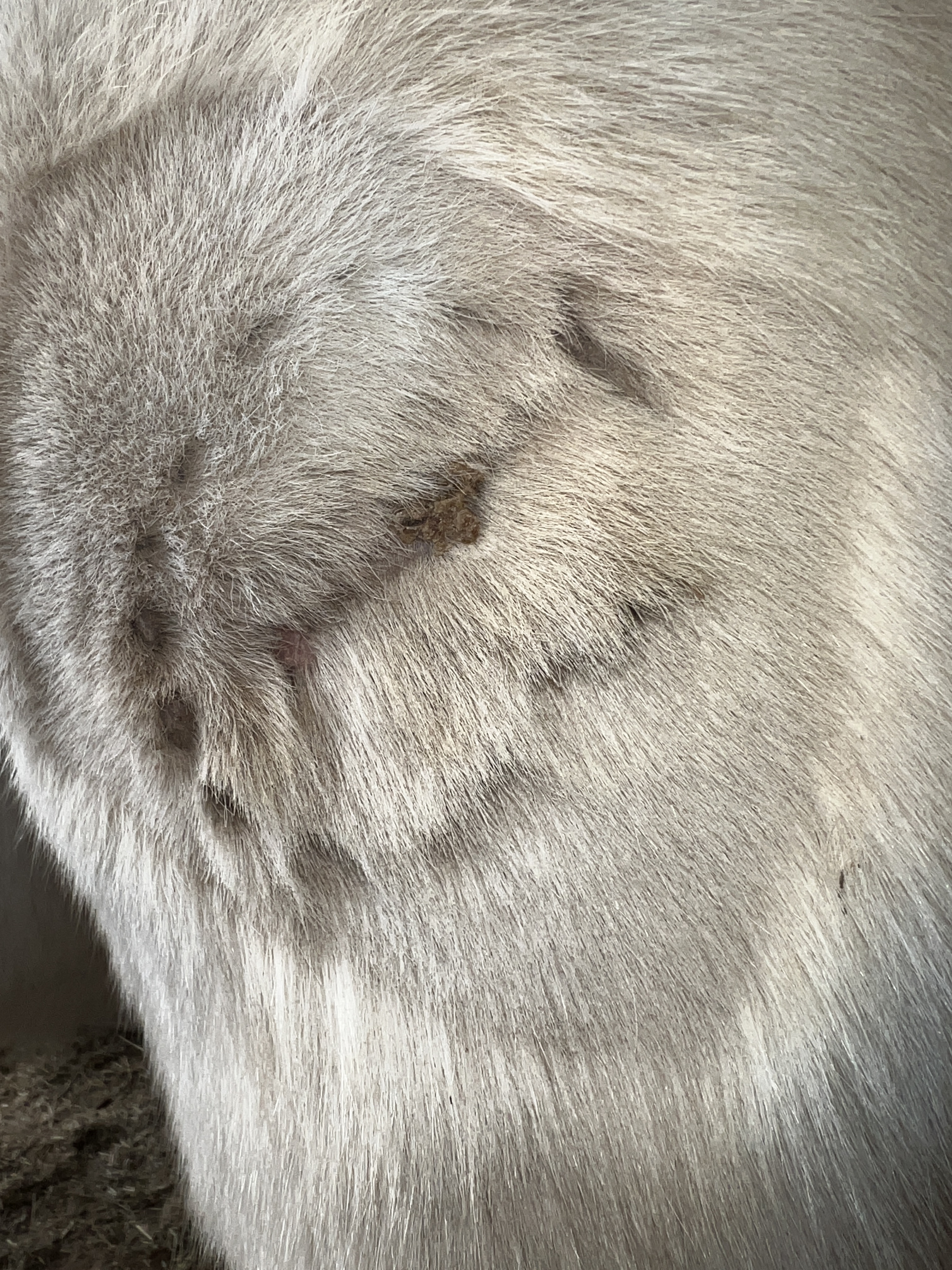
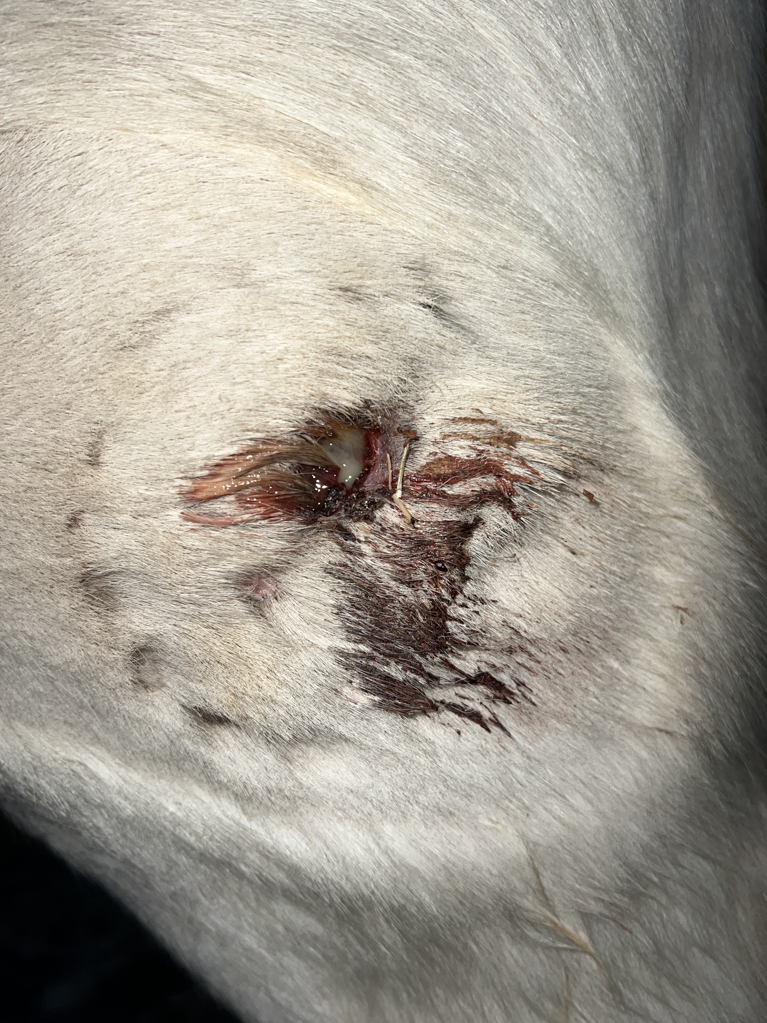
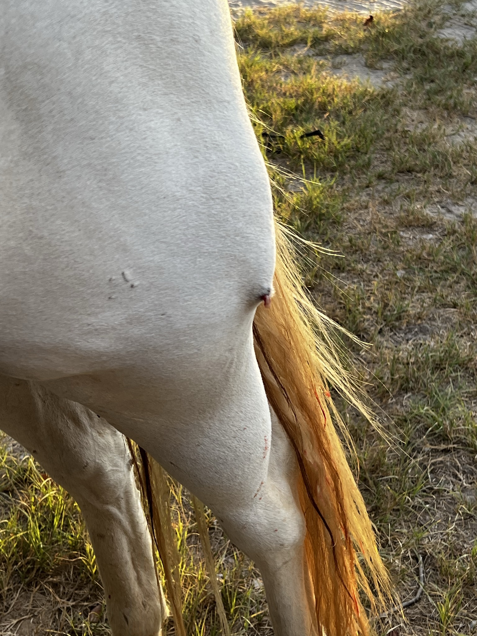
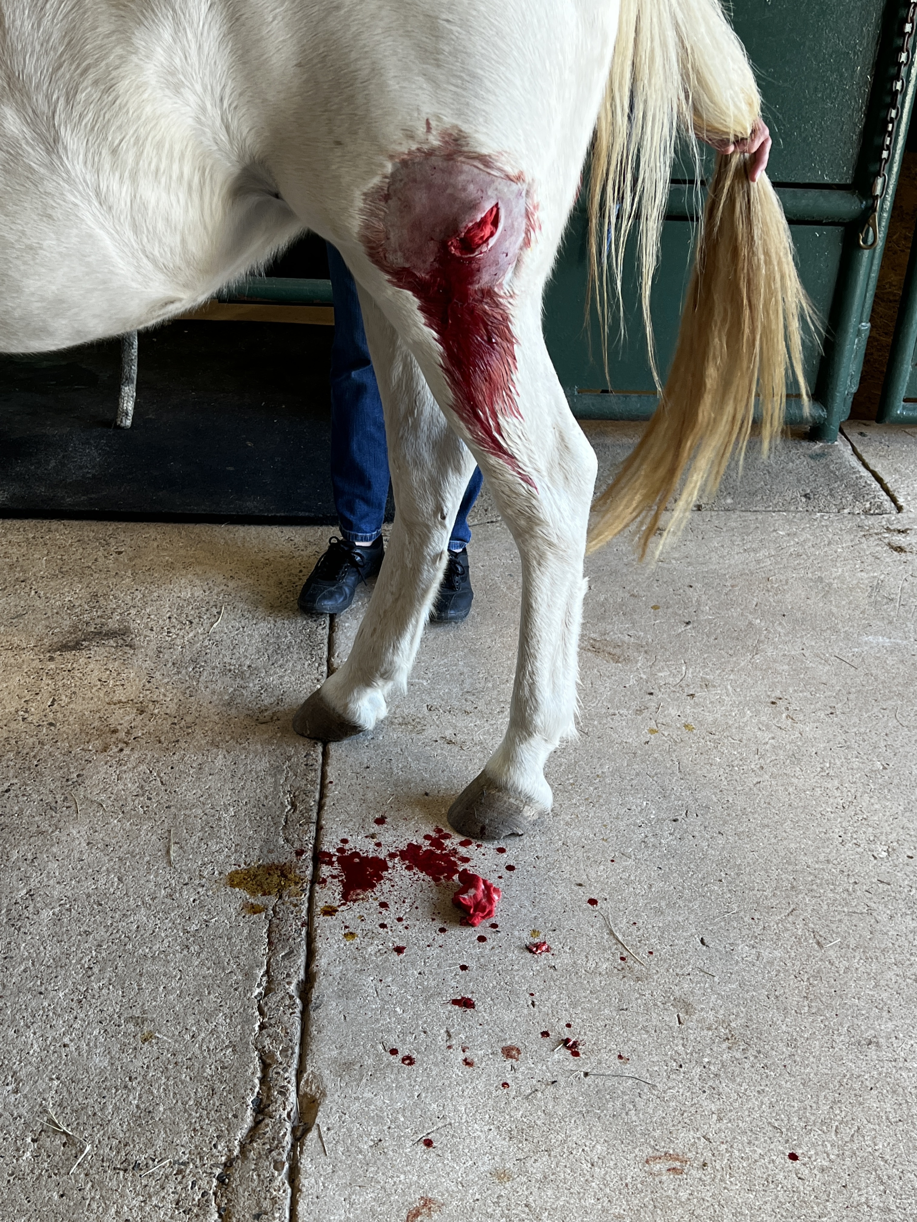
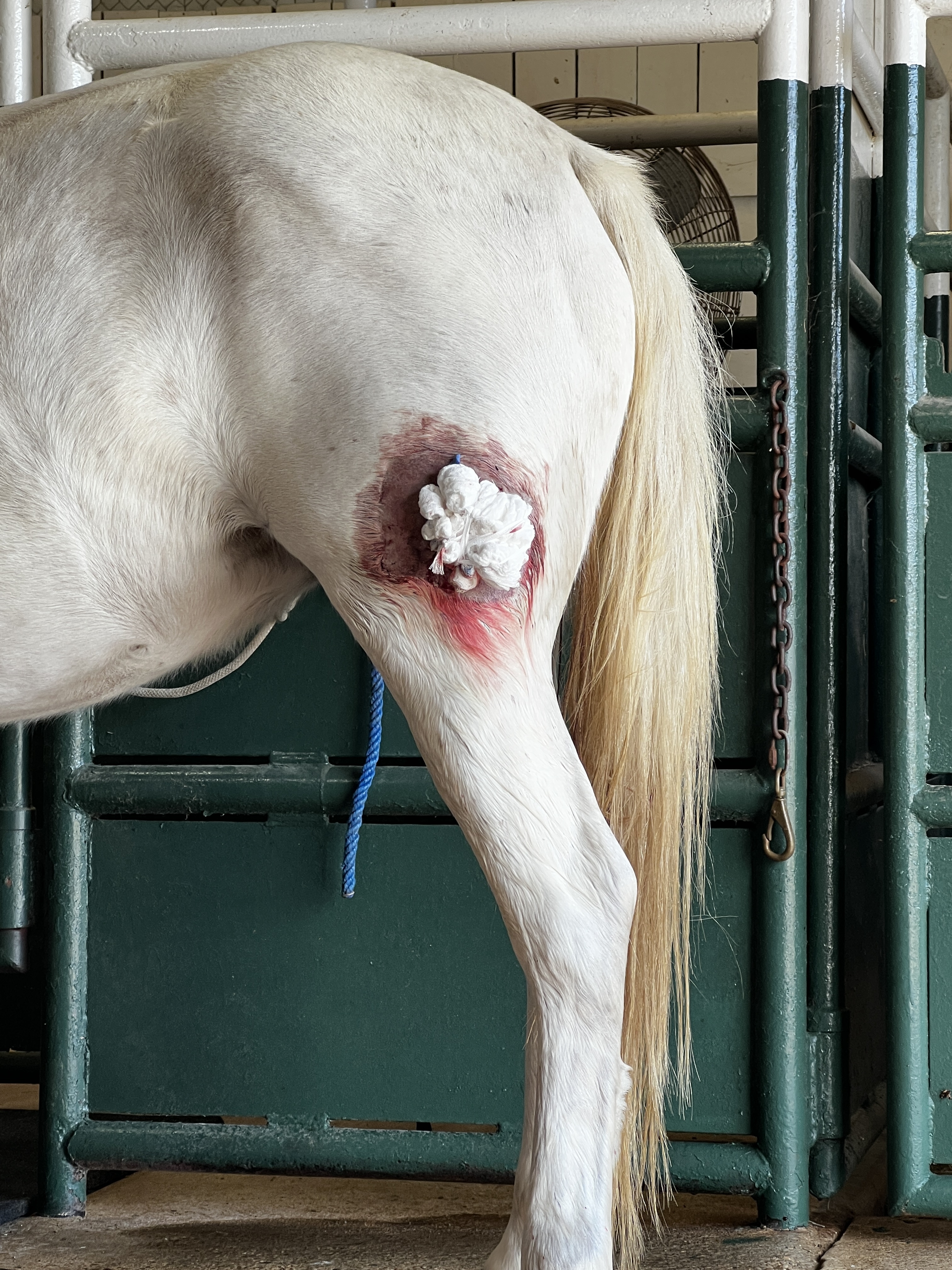
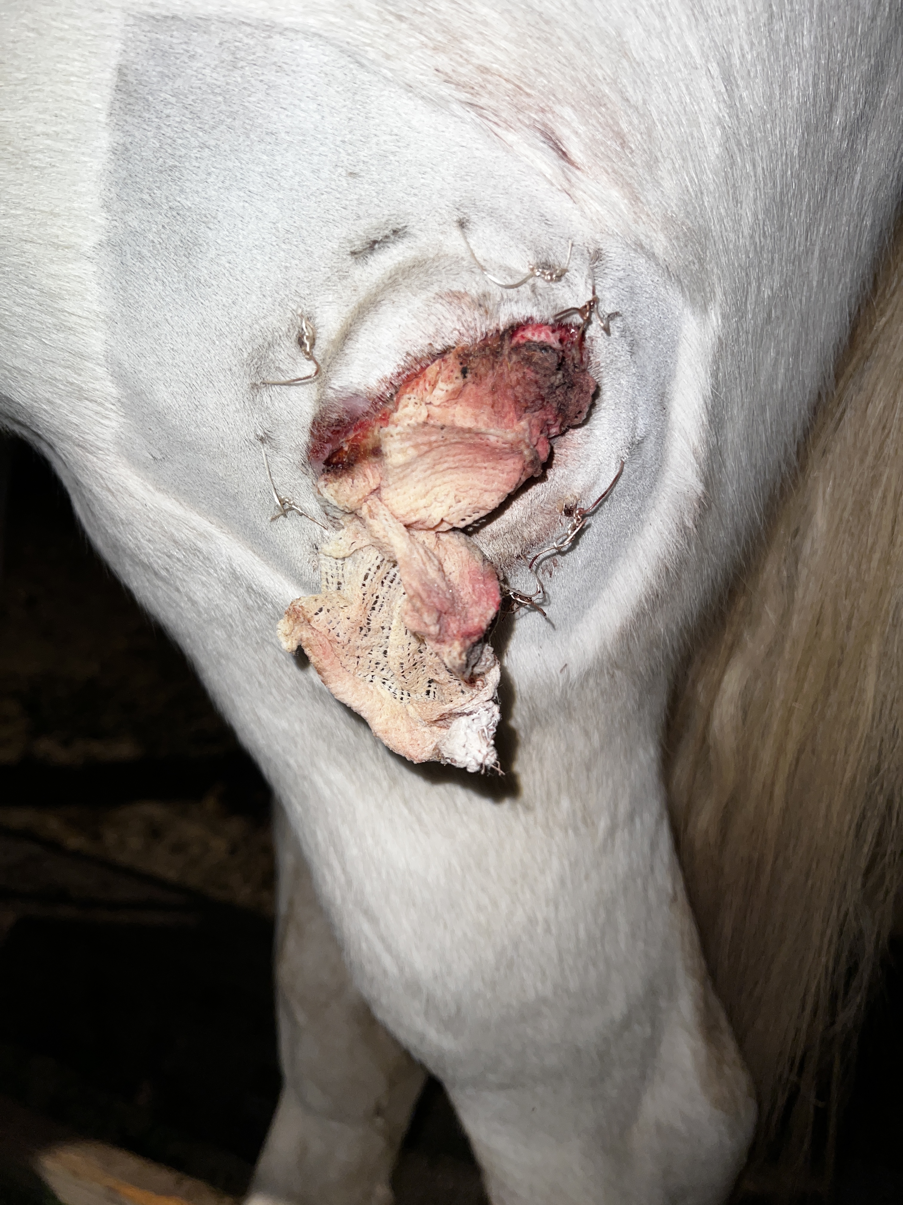
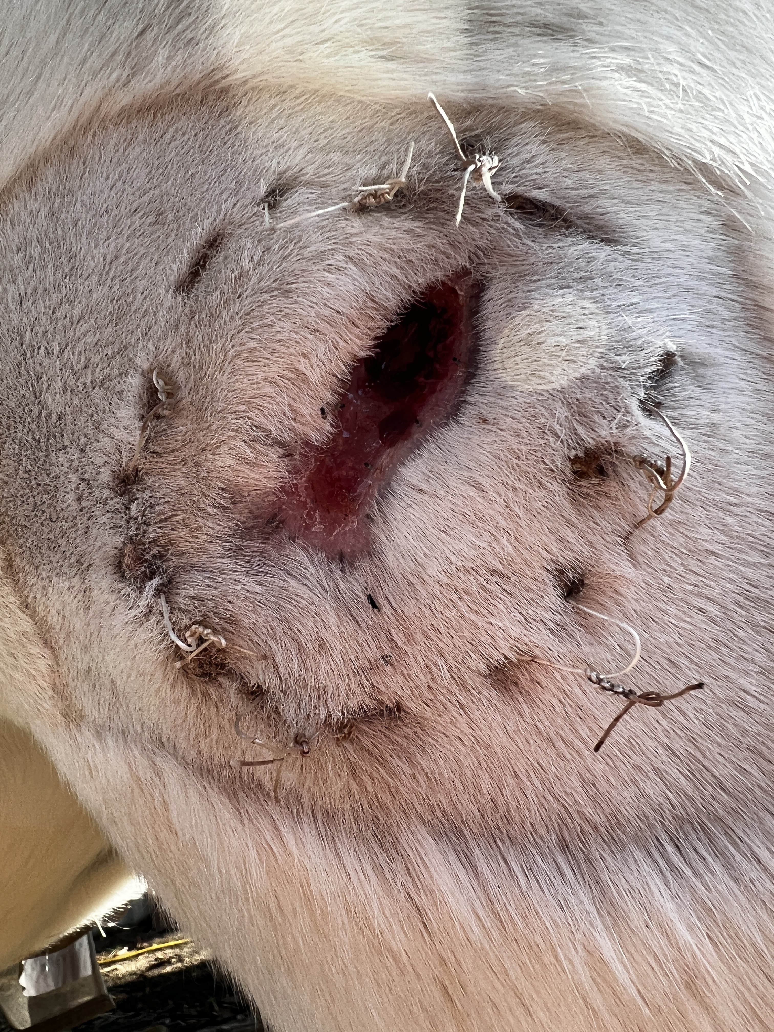
This pony mare developed an abscess in a period of a few days on July 27th. There was no broken skin previously. I rode her and she suddenly couldn’t bring her left hind up and forward to step over a pole, 3 days later a huge abscess erupted right before my eyes as I brought her in from pasture. Kept it clean let it drain, didn’t heal so we did a round of uniprim, still didn’t heal. It would heal to a pin point but still drain and have a hard feeling behind it. Beginning of October the vet ultrasounded it and found nothing, X-rays showed nothing, no sequestrum, she debrided 8 inches into the “abscess” pulling out dead scar tissue until she couldn’t reach anymore. She was on doxycycline twice a day for a month and metronidazole 3 times a day for a month. Did bandage changes every 3 days until it closed. It was officially closed and I removed the stitches on November 17th and here she is with this damn thing again! It’s all hard around it. In the center of the exposed tissue is a grey spot. I cleaned it with betadine and put curicyn clay on it. Not sure what else this can be but the next step is the vet putting her under general and slicing her leg open entirely and even then she said it’s no guarantee that we will find anything. I’m beginning to wonder if this is cancer, she already has melanoma under the dock of her tail. She’s a 2008 Connemara pony. If it is cancer I wonder what the treatment and prognosis is or if euthanasia would be the best option.

!
[image|375x500]

I know with humans, if the sac is not removed it can come back, can your vet try to remove it, and have it biopsied? What about an ultrasound or some sort of imaging to see if something is stuck up in there that needs to come out?

If you read the post you’ll see it was debrided and everything was removed as far as a “sac” and old scar tissue tracts. It was also ultrasounded and xrayed.

1 Like

Wow! I’d also want a culture at this point, and I’d push hard for some tissue biopsies, sent to whatever the best lab in the country is for that stuff.

5 Likes

I just meant could they look again like when it drains or if they have biopsied anything? Either way I’m sorry and I hope it clears up for her.

1 Like

I think that what was being offered is that, while we know how incredibly frustrating this is for you and your girl, the abscess wouldn’t come back unless whatever was causing it to begin with was still in there. It would be awfully odd for something new to cause an abscess in the same spot, right?

Good luck with this- it is a challenge.

9 Likes

There could be something in there, however the vet cut out the entire abscess the first time, opened the wound and dug around 8 inches using her fingers and various tools and found nothing. The tissue was healthy when she cleaned everything out. Nothing on ultrasound or x ray. No original entry point for an abscess to occur. There was no broken skin there.
We originally hypothesized that maybe she tore a muscle, we did some magnawave and 3 days later this beauty erupted. About 6 months prior to this erupting in July she was kicked by a barefoot mare, didn’t break the skin but was a good goose egg. She wasn’t lame from it. If it’s not an abscess, if it’s not cancer, could it be an old hematoma? The tract the vet followed went up between the 11 and 12 o’clock 8 inches above the entry. The tract was made up of “deranged” tissue and she removed it as she went along. Every bandage change there was no tract formation and no deranged tissue. It was all healthy tissue as it closed. Even this reappearance is so superficial. Like it’s just hard in the skin and a little bit behind it. Just like how it healed the first time. But she cut alllll that out so I don’t know why it’s back. The drawing clay is pulling out a lot of yellow infection. If it really is a foreign body I’m wondering if the body will be more effective at getting rid of it then cutting her open again will be. She was on heavy duty antibiotics for aerobic and anaerobic bacteria for a month!

Just because broad spectrums were used doesn’t mean that was the right tool for the job. It’s better to culture and pick an agent that’s very specifically effective against whatever bacteria this is. Get a culture & sensitivity.

A biopsy will tell you if you’re dealing with cancer, or an autoimmune issue, or something else abnormal in the cells themselves.

9 Likes

All 3 antibiotics used stopped the infectious process. It doesn’t matter if you’re using the correct specific antibiotic that a bacteria is sensitive to if the root cause of the issue isn’t addressed and it’s not just a bacterial infection. This is now a 3 time recurrent sore. If it really is a foreign body then antibiotics aren’t going to fix it when it needs to be removed either by the body’s infectious process or physically (which we’ve attempted). My next step is a biopsy. I wanted to know if this looks like a familiar cancer to anyone and if so, what was the prognosis and treatment like. Or there’s the off chance that this is a traumatic hematoma with some muscle damage on the inside. Or it could still be a foreign body and I guess I will just rinse and repeat this until it works it’s way out. :woman_shrugging:

1 Like

I’m sorry you’re struggling with this challenge. Like many others who’ve had gray horses, I’ve done the melanoma thing. Years ago I had a gray gelding that had external melanomas that eventually created drainage “tracts” that coursed through muscle tissue, eventually erupting on the outside. I am NOT saying that’s what this is, but it is curious that your vet did so much exploration, seemed to find healthy tissue, yet the site has re-opened again.

On the other hand, perhaps it’s still a smoldering infection that just won’t die, and now it’s become a chronic condition. Intravenous antibiotics vs. the oral route, may do the trick. But I would think that going back in there and taking numerous biopsies and culture and sensitivity studies, would make sense. It will help identify exactly what it is you’re fighting.

Here’s a link to the Merck manual on muscle abscesses.
https://www.merckvetmanual.com/musculoskeletal-system/myopathies-in-horses/infectious-myopathies-in-horses#:~:text=Staphylococcus%20aureus%2C%20Streptococcus%20equi%2C%20and,or%20local%20spread%20of%20infection.

1 Like

Egads! No further advice, but plenty of jingles!

Having gone multiple rounds with a complicated infection quite recently, yes, a culture and sensitivity is important. It’s entirely possible to have a small amount of infectious material that is not completely addressed by whatever antibiotics you’ve used, that will then regrow once you withdraw treatment, because you think you’ve dealt with it.

Frankly, it’s pretty shocking you haven’t gotten one before now. It’s not a complicated or particularly costly thing to do, and allows you to actually target your therapy.

As someone who has been down this path, what you think you’re dealing with and what you’re actually dealing with can be pretty different. And what you think you should be using, antibiotic-wise, and what you should actually be using can be pretty different, too.

There’s literally zero reason to skip a c/s at this point. What have you got to lose?

7 Likes

totally agree that this is culture sensitivity time AND pathology needed.

foreign plant tissue like a bit of thorn or stick will not be viewable on x-ray and could be very hard to palpate during debridement. It can be terribly hard to track this down

Melanoma needs to be highly considered. They are not always black but can become gray or red pink which certainly could mimic infected tissues .

2 Likes

My abscess knowledge is human related, but oral antibiotics usually aren’t great for abscesses. They don’t have a great blood supply because the immune system will try to surround them and cut them off from the rest of the body to prevent a systemic infection. So even if you are giving very strong antibiotics, it doesn’t necessarily mean the body is allowing them to reach the abscess.

In human medicine they sometimes will irrigate the old abscess site with antibiotics.

2 Likes

We just did this with a lesson horse who had been kicked. My vote is foreign body.

Presented with a kick wound. Wound was minor, horse was sound. Used him in a lesson the next day. The following day it BLEW UP. Took him to hospital and stayed overnight, then following process took a month to get him back to light work. Vet said he sees it often. Kick causes tissues to separate, inviting foreign bodies which would normally just attach to hair/skin to come in and migrate deep. In our case we were LUCKY to find a teensy piece of wood. I’m talking the tip of a needle teensy. The fact that I used him in exercise likely further invited the foreign object to continue penetrating due to movement (near the knee).

Good luck OP. I do think you’ll get to then bottom of this!

2 Likes

I had a colt, many years ago, who developed a fistulous track on the lateral aspect of his front left fetlock. It was draining intermittently. We had no idea what was causing it. I took him over to the local Veterinary surgeon’s hospital (Phylliss Lose) and she did surgery, flushed the tract, and found the tiniest little bone shard in there. I figure that his Mama may have stepped on him, or something, and there was just this tiny microscopic splinter that caused the issue. He healed up, and it disappeared.

I think that point is that there can be something in there that is tiny, but is lodged in there, and just keeps re-infecting the tissue. It could be something higher up the drain field, and the infection is inoculated by the foreign body, and lands where you are seeing it. Presto! It’s baaaaackkk!

3 Likes

When I was 5 I had a little accident and eight fingers, all but the thumbs, were crushed, some more than others.
They healed fine, but 10 years later, the little finger on the left started getting a bump, that kept growing and growing.
Dr finally cut it out, sent it to the lab and its seed was a tiny sliver of bone, probably from that accident 10 years ago, that had encapsulated and finally traveled to the outside and may have eventually popped out.

Such happens, could be what is happening here from the kick, but in a grey horse and with infection having set in, maybe a better/research horse hospital could handle that kind of search surgery and lab work better at this point?

1 Like

We will definitely do a C&S. I’m not sure why we initially didn’t do one. Vet seemed confident in her choice of antibiotics. I’d hate to keep dumping money into a mare I intended to sell but I wonder if a CT scan would be able to identify where exactly we need to treat. Especially if it is a foreign body maybe it can be extracted more easily and then treated with antibiotic flushing/systemic abx.

1 Like

That’s what a sequestrum is called and X-rays were negative. :frowning:

The tissue I was looking at when I initially cleaned up this reopening the wound was very strange looking I have no clue why I didn’t take a picture. There is a grey spot right in the middle. I’m going to scrub it again today and see if I can’t get a good visual to show y’all.